A pair of oval glands hanging outside the abdomen so that they stay 2–4 degrees cooler than the body core
Testis — sperm production works best below core temperature.
Anatomy · Glands & the immune system
The male gonad — one on each side of the scrotum
Reproductive and endocrine systems · scrotum
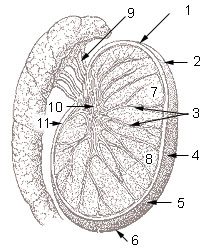
One of a pair of oval glands hanging in the scrotum outside the abdomen, where it is kept a few degrees cooler than the body core. Each testis is divided into lobules packed with coiled seminiferous tubules, where sperm are made, and it also makes the hormone testosterone. Sperm collect in the rete testis and pass through the efferent ductules into the epididymis, which sits along its back, and then into the vas deferens.
A pair of oval glands hanging outside the abdomen so that they stay 2–4 degrees cooler than the body core
Testis — sperm production works best below core temperature.
Inside a white fibrous capsule, several hundred lobules hold the coiled tubules in which sperm are made
Testis — the seminiferous tubules, lined by Sertoli cells.
Cells between the tubules, described by Franz Leydig in 1850, make the male sex hormone (1850)
Testis — Leydig (interstitial) cells make testosterone.
A web of veins wraps its artery in the cord so blood is cooled on the way in
Testis — the pampiniform plexus works as a countercurrent heat exchanger.
Its lymph drains to nodes beside the aorta rather than the groin, which matters in cancer staging
Testis — para-aortic nodes, because it began life high in the abdomen.
It begins life in the abdomen and travels down the inguinal canal into a pouch late in fetal life, around the seventh month
Testis — failure to descend is cryptorchidism.
Its Latin name is also the Latin word for 'witness'
Testis — the origin of the word 'testify' is often linked, though the etymology is uncertain.