Leaves the skull through a foramen shared with cranial nerves IX, X and XI, as the continuation of the sigmoid sinus
Jugular vein — the internal jugular begins at the jugular foramen.
Anatomy · Heart & blood vessels
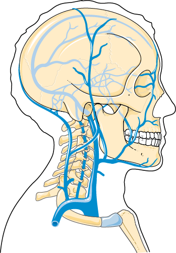
Main venous drainage of the head and neck
Neck · carotid sheath and superficial neck
The jugular veins drain blood from the head and neck. The internal jugular vein, the larger, leaves the skull through the jugular foramen and runs down within the carotid sheath, lateral to the carotid artery, to join the subclavian vein and form the brachiocephalic vein. The external jugular vein runs superficially across sternocleidomastoid and is visible under the skin.
Leaves the skull through a foramen shared with cranial nerves IX, X and XI, as the continuation of the sigmoid sinus
Jugular vein — the internal jugular begins at the jugular foramen.
Runs down the neck in the carotid sheath, lateral to the common carotid artery
Jugular vein — the internal jugular vein.
It joins the subclavian vein behind the sternal end of the clavicle to form the brachiocephalic vein
Jugular vein — the venous angle.
Its visible pulsation in the neck, measured from the sternal angle, indicates pressure in the right atrium
Jugular vein — the jugular venous pressure (JVP).
A superficial vein that can be seen crossing the sternocleidomastoid when someone coughs or strains
Jugular vein — the external jugular vein.
A common site for inserting a central venous catheter under ultrasound guidance, on the right side by preference
Jugular vein — internal jugular cannulation.
The thoracic duct on the left and the right lymphatic duct on the right empty at the junction where this vein meets the subclavian
Jugular vein — the jugulo-subclavian junction.