🪴 GoDeep
Search
← University Challenge
Portrait of Circle of Willis

Anatomy · Heart & blood vessels

Circle of Willis

Described by Thomas Willis, 1664

Base of the brain · arterial anastomosis

A ring of arteries at the base of the brain that joins the two internal carotid arteries (the anterior circulation) to the basilar artery (the posterior circulation, fed by the vertebral arteries). It lets blood reach any part of the brain by an alternative route if one supplying vessel is narrowed or blocked. It is named after the English physician Thomas Willis, who described it in his Cerebri Anatome of 1664.

How to spot them

  • ▸A polygon of vessel segments: the anterior communicating artery, two anterior cerebral arteries, two internal carotid arteries, two posterior communicating arteries and two posterior cerebral arteries
  • ▸Lies in the interpeduncular cistern in the subarachnoid space, around the optic chiasm and pituitary stalk
  • ▸Fed by the two internal carotid arteries in front and the basilar artery (from the two vertebral arteries) behind
  • ▸Anatomical variants (a small or missing communicating artery) are found in a large proportion of people

What to know

A ring of arteries at the base of the brain, linking the carotid and vertebrobasilar systems

Circle of Willis — anastomosis of the anterior and posterior circulation.
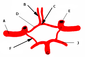
Named after a seventeenth-century English physician who described it, with a drawing by Christopher Wren, in a book on the anatomy of the brain

Circle of Willis — Thomas Willis, Cerebri Anatome (1664), illustrated by Sir Christopher Wren.

Its front is closed by a short artery joining the two anterior cerebral arteries

Circle of Willis — the anterior communicating artery.

Its back is closed by two small arteries, each joining an internal carotid to a posterior cerebral artery

Circle of Willis — the posterior communicating arteries.

Berry aneurysms, whose rupture causes subarachnoid haemorrhage, form most often at junctions in this ring

Circle of Willis — commonly the anterior communicating artery junction.

Provides collateral flow so that blockage of one carotid may cause no symptoms

Circle of Willis.

Surrounds the optic chiasm and the stalk of the pituitary gland

Circle of Willis — in the interpeduncular cistern.

Key ideas

  • Anastomosis, not a full circle in most people: it is complete in only a minority to about half of people, depending on the series studied; missing or small posterior communicating arteries are commonest.
  • The internal carotid arteries give the anterior part; the basilar artery, from the union of the two vertebral arteries, divides into the two posterior cerebral arteries.

Quick-fire facts

  • •Named after Thomas Willis (1621–1675), who described it in Cerebri Anatome (1664); Christopher Wren made the drawings.
  • •Composed of the anterior communicating artery, the two anterior cerebral arteries (A1 segments), the two internal carotid arteries, the two posterior communicating arteries and the two posterior cerebral arteries (P1 segments).
  • •The brain uses about 20% of the body's oxygen and receives about 15% of cardiac output.
  • •Anterior communicating artery aneurysms are the commonest site of berry aneurysm.
  • •The basilar artery is formed by the union of the two vertebral arteries at the pontomedullary junction.
  • •Complete polygons are found in roughly 20–50% of people depending on the study.
  • •Willis also coined the term 'neurology' and described the eleventh cranial nerve, the accessory nerve.
  • •A ruptured berry aneurysm produces a sudden 'thunderclap' headache, often described as the worst of the patient's life.

Drill it — 7 cards

1 / 7new card

Sources